Easy access to products and protocols for research use only in the identification of 2019-nCoV based on Centers for Disease Control and Prevention (CDC) recommendations
So much has changed during this unprecedented time, except your ability to count on Avantor. We continue to set science in motion to create a better world by providing you with the right solutions to keep moving forward.
Our solutions, developed with you as our focus, are crafted by our team and network of professionals with advanced degrees in science, quality control, engineering, manufacturing and industry experience.
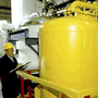
Avantor supports end-to-end fluid management solutions – including peristaltic pumps and aseptic fluid transfer solutions – that are reliable and customer-centric, helping bioprocessing manufacturers meet their research and production goals.
A strong, vibrant research and development group is the lifeblood of all industries. VWR will support you from the latest life science products to the guaranteed purity of organic building blocks...
VWR is ready to support your production facility with reliable access to raw materials and essential supplies. We can also help you increase productivity...
VWR is proud of our years of experience providing choice and excellent service to the Industrial market from Food & Beverage, Petrochemical, Environmental Testing, Waste Water, Cosmetics, Consumer Goods, Agriculture and more...
VWR is your complete source for workplace supplies. Binders, calendars, pens, cleaning and sanitation supplies, and office equipment are just some of the essential products we offer...
New Avantor® J.T.Baker® premium conductive and non-conductive robotic tips deliver superior quality and reliable performance for results you can trust.
Avantor Services provides a wide range of specialized services and digital solutions to help you solve complex challenges.
We’ve built our reputation on consistent, comprehensive mastery of day-to-day operations, allowing lab, clinical, and production environments to focus their high-value resources on core scientific priorities.
As our customers’ needs have evolved, so have our capabilities. We have become experts in scientific operations, improving performance with sophisticated solutions and providing guidance on best practices.
You can select and customize services for peak efficiency, quality, and accelerated innovation.
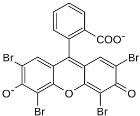
Microbiology stains are used to detect and identify microorganisms that would otherwise be difficult to find in clinical specimens. Simple positively or negatively charged dyes create a fast discovery. The premixed solutions artificially color to help observe movement, structure, and characteristics of live or preserved specimens. Differentiating microorganisms based on properties, color combination procedures like the Gram stain or acid-fast techniques require microbiology stains.
Description:
Mayer Mucicarmine Method, For mucosubstances stains, Note: rose color due to carmin staining will be obscured if sections are overstained with Weigert's Hematoxylin and/or Metanil Yellow solution.
Description:
Taylor's Method for Gram Positive and Gram Negative Bacteria, Stain results - Gram-Positive Organism: Blue to Blue-Black, Gram Negative Organism: Bright Red, Nuclei: Brownish Red, Erythrocytes: Red to Yellow-Green
Description:
Mayer Mucicarmine Method, For mucosubstances stains, Note: rose color due to carmin staining will be obscured if sections are overstained with Weigert's Hematoxylin and/or Metanil Yellow solution.
Description:
Mayer Mucicarmine Method, For mucosubstances stains, Note: rose color due to carmin staining will be obscured if sections are overstained with Weigert's Hematoxylin and/or Metanil Yellow solution.
Description:
Taylor's Method for Gram Positive and Gram Negative Bacteria, Stain results - Gram-Positive Organism: Blue to Blue-Black, Gram Negative Organism: Bright Red, Nuclei: Brownish Red, Erythrocytes: Red to Yellow-Green
Description:
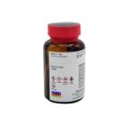
Gridley's Method for Fungi For stain fungal mycelia, conidia, morphological detail of yeast forms and hyphae. Stain results - Mycelia, Conidia: Deep Purple Elastic Tissue & Mucin Background: Yellow
Description:
Gridley's Method for Fungi For stain fungal mycelia, conidia, morphological detail of yeast forms and hyphae. Stain results - Mycelia, Conidia: Deep Purple Elastic Tissue & Mucin Background: Yellow