


You Searched For:
519 results were found
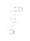
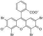
Histology Stains
|
|||||||||||||||||||||||||||||||||||||||||||||||||||||||||||||||||||||||||||||||||||||||||||||||||||||||||||||||||||||||||||||||||||||||||||||||||||||||||||||||||||||||||||||||||||||||||||||||||||||||||||||||||||||||||||||||||||||||||||||||||||||||||||||||||||||||||||||||||||||||||||||||||||||||||||||||||||||||||||||||||||||||||||||||||||||||||||||||||||||||||||||||||||||||||||||||||||||||||||||||||||||||||||||||||||||||||||||||||||||||||||||||||||||||||||||||||||||||||||||||||||||||||||||||||||||||||||||||||||||||||||||||||||||||||||||||||||||||||||||||||||||||||||||||||||||||||||||||||||||||||||||||||||||||||||||||
![]()
Avantor Services provides a wide range of specialized services and digital solutions to help you solve complex challenges.
We’ve built our reputation on consistent, comprehensive mastery of day-to-day operations, allowing lab, clinical, and production environments to focus their high-value resources on core scientific priorities.
As our customers’ needs have evolved, so have our capabilities. We have become experts in scientific operations, improving performance with sophisticated solutions and providing guidance on best practices.
You can select and customize services for peak efficiency, quality, and accelerated innovation.
For more information, call 1.888.793.2300.
![]()
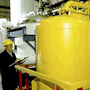
The Equipment & Instrument Service Team’s focus is to provide comprehensive service solutions for our customers. We provide validation, calibration, preventative maintenance, and extended warranties on all equipment & instruments in and around the laboratory.
For additional information call 1.888.793.2300 or email us at equipmentservices.us@avantorsciences.com
Avantor® is a leading life science tools company and global provider of mission-critical products and services to customers in the life sciences and advanced technology industries. From discovery to delivery, we work side-by-side with scientists around the world to enable breakthroughs in medicine, healthcare, and technology. Our portfolio is used in virtually every stage of the most important research, development and production activities at more than 300,000 customer locations in 180 countries. For more information, visit avantorsciences.com and find us on LinkedIn, X (Twitter) and Facebook . © 2024 VWR International, LLC. All rights reserved. | |||||||||||||||||||||||||||||||||||||||||||||||||||||||||||||||||||||||||||||||||||||||||||||||||||||||||||||||||||||||||||||||||||||||||||||||||||||||||||||||||||||||||||||||||||||||||||||||||||||||||||||||||||||||||||||||||||||||||||||||||||||||||||||||||||||||||||||||||||||||||||||||||||||||||||||||||||||||||||||||||||||||||||||||||||||||||||||||||||||||||||||||||||||||||||||||||||||||||||||||||||||||||||||||||||||||||||||||||||||||||||||||||||||||||||||||||||||||||||||||||||||||||||||||||||||||||||||||||||||||||||||||||||||||||||||||||||||||||||||||||||||||||||||||||||||||||||||||||||||||||||||||||||||||||||||||||||||