Easy access to products and protocols for research use only in the identification of 2019-nCoV based on Centers for Disease Control and Prevention (CDC) recommendations
So much has changed during this unprecedented time, except your ability to count on Avantor. We continue to set science in motion to create a better world by providing you with the right solutions to keep moving forward.
Our solutions, developed with you as our focus, are crafted by our team and network of professionals with advanced degrees in science, quality control, engineering, manufacturing and industry experience.
Avantor supports end-to-end fluid management solutions – including peristaltic pumps and aseptic fluid transfer solutions – that are reliable and customer-centric, helping bioprocessing manufacturers meet their research and production goals.
A strong, vibrant research and development group is the lifeblood of all industries. VWR will support you from the latest life science products to the guaranteed purity of organic building blocks...
VWR is ready to support your production facility with reliable access to raw materials and essential supplies. We can also help you increase productivity...
VWR is proud of our years of experience providing choice and excellent service to the Industrial market from Food & Beverage, Petrochemical, Environmental Testing, Waste Water, Cosmetics, Consumer Goods, Agriculture and more...
VWR is your complete source for workplace supplies. Binders, calendars, pens, cleaning and sanitation supplies, and office equipment are just some of the essential products we offer...
New Avantor® J.T.Baker® premium conductive and non-conductive robotic tips deliver superior quality and reliable performance for results you can trust.
Avantor Services provides a wide range of specialized services and digital solutions to help you solve complex challenges.
We’ve built our reputation on consistent, comprehensive mastery of day-to-day operations, allowing lab, clinical, and production environments to focus their high-value resources on core scientific priorities.
As our customers’ needs have evolved, so have our capabilities. We have become experts in scientific operations, improving performance with sophisticated solutions and providing guidance on best practices.
You can select and customize services for peak efficiency, quality, and accelerated innovation.
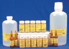
Overcome normal optical limitations in microscopic studies by highlighting specific structures with stains and reagents and then protect samples with fixatives. The fast-acting dyes create strong contrasts between cell components for easier detection or analyses. Sensitive attractions to certain organelle or cellular components differ among offered stains, allowing precise testing modifications. Flexible in use, ready-to-use stains may be handled manually or in conjunction with automatic equipment.
Description:
excellent adhesion to metals, glass, and ceramic, use as a water soluble temporary bond, for precise high purity work, leaves no residue after dissolving, does not clog the diamond wheel,
Description:
Ready to use, protects cellular structure, minimal interference with subsequent staining, for use with human and animal calcified tissue, bone, cartilage and other hard specimens
Description:
A Combined Hematoxylin and Eosin/Methenamine Silver Stain for the Histological Diagnosis of Fungi in Tissue Sections For bacterial, fungal stains. Stain results - Organism: Blue-Black Background: Rose
Description:
A Combined Hematoxylin and Eosin/Methenamine Silver Stain for the Histological Diagnosis of Fungi in Tissue Sections For bacterial, fungal stains. Stain results - Organism: Blue-Black Background: Rose
Description:
A thermoplastic adhesive for mounting powder specimens and small particles for SEM. Does not contain any solvents and is stable in high vacuum. It is not sticky at room temperature but becomes adhesive at 40[degree]C and melts at 120[degree]C.
Description:
Modified Steiner - Using Chapman's Modification For bacterial stain. Stain results - Spirochetes,Donovan Bodies, General Bacteria,Legionairies Disease Bacteria: Dark Brown or Black Background: Bright Yellow
Description:
Laqueur's Method for Alcoholic Hyalin Lacqueur (1950), For mallory bodies, erythrocytes, bile pigment and proteinaceous, material in liver, Stainresults - Mallory Bodies: Bright Red, Erythrocytes: Red, Cytoplasm: Pale Brown
Description:
Thomas Method for Malarial Parasites, Thomas (1953), Stain results - Nuclei: Blue, Plasma Cell Cytoplasm: Blue, Malarial Parasites: Blue, Erythrocytes: Pink, Other Tissue Elements: Shades of Rose to Red
Description:
Giemsa Thick Film Stain, Lillie (1965); Barber and Komp (1929), Stain malarial parasites, Stain results - Malarial Parasites: Clear Red Chromatin, Cytoplasm: Clear Blue, Red Corpuscles: Not seen due to lacking of
Description:
Prussian Blue Method for Hemosiderin, For the staining of deposits of hemosiderin, Stain results - Hemosiderin: Blue or Green, Nuclei: Red, Background: Pink
Description:
Carstairs Method for Fibrin and Platelets, For the staining of fibrin and platelets, Stain results - 48 hours or more: Fibrin: Bright Red, Platelets Gray blue to navy, Collagen: Bright Blue, Muscle: Red
Description:
Alizarin Red S and Toluidine Blue O, Williams (1941); Dawson (1926), Distinction between bone and cartilage in mammalian embryos, Stain results - Soft Tissues: Transparent, Osseous Tissues: Deep Red, Cartilage: Dark Blue"
Description:
McManus' Method (PAS) for Glycogen, McManus (1948), Stain results - Nuclei: Blue, Fungi: Red, Background, when Light Green is used as the counter-stain: Green, pale
Description:
Jones Method for Kidney, Silver Method, For the staining of basement membranes, reticulum fibers, collagen, and nuclei, Stain results - Basement membranes, reticulum fibers: Black, Nuclei: Blue, Cytoplasm, collagen
Description:
designed as a masking material in jet polishing applications, chemically inert, resistant to etching solutions (hydrofluoric acid, perchloric acid and acetic acid), can be easily removed with hydrocarbon or chlorinated solvents, M
Description:
Alizarin Red S and Toluidine Blue O, Williams (1941); Dawson (1926), Distinction between bone and cartilage in mammalian embryos, Stain results - Soft Tissues: Transparent, Osseous Tissues: Deep Red, Cartilage: Dark Blue
Description:
Lillie's Method for Iron, For the staining of ferric and ferrous iron, Stain results - Ferric Iron: Dark Prussian Blue, Ferrous Iron: Dark Turnbull's Blue, Background: Light Red