Easy access to products and protocols for research use only in the identification of 2019-nCoV based on Centers for Disease Control and Prevention (CDC) recommendations
So much has changed during this unprecedented time, except your ability to count on Avantor. We continue to set science in motion to create a better world by providing you with the right solutions to keep moving forward.
Our solutions, developed with you as our focus, are crafted by our team and network of professionals with advanced degrees in science, quality control, engineering, manufacturing and industry experience.
Avantor supports end-to-end fluid management solutions – including peristaltic pumps and aseptic fluid transfer solutions – that are reliable and customer-centric, helping bioprocessing manufacturers meet their research and production goals.
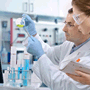
A strong, vibrant research and development group is the lifeblood of all industries. VWR will support you from the latest life science products to the guaranteed purity of organic building blocks...
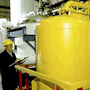
VWR is ready to support your production facility with reliable access to raw materials and essential supplies. We can also help you increase productivity...
VWR is proud of our years of experience providing choice and excellent service to the Industrial market from Food & Beverage, Petrochemical, Environmental Testing, Waste Water, Cosmetics, Consumer Goods, Agriculture and more...
VWR is your complete source for workplace supplies. Binders, calendars, pens, cleaning and sanitation supplies, and office equipment are just some of the essential products we offer...
New Avantor® J.T.Baker® premium conductive and non-conductive robotic tips deliver superior quality and reliable performance for results you can trust.
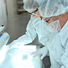
Avantor Services provides a wide range of specialized services and digital solutions to help you solve complex challenges.
We’ve built our reputation on consistent, comprehensive mastery of day-to-day operations, allowing lab, clinical, and production environments to focus their high-value resources on core scientific priorities.
As our customers’ needs have evolved, so have our capabilities. We have become experts in scientific operations, improving performance with sophisticated solutions and providing guidance on best practices.
You can select and customize services for peak efficiency, quality, and accelerated innovation.
The Equipment & Instrument Service Team’s focus is to provide comprehensive service solutions for our customers. We provide validation, calibration, preventative maintenance, and extended warranties on all equipment & instruments in and around the laboratory.
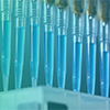
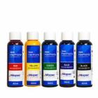
Used for biological or medical research, histology stains and reagents support easy microscopic anatomy analysis. Powder or liquid form stains effectively highlight plant or animal cell and tissue components. When used in conjunction with microscopic equipment, the premium and environmentally friendly chemicals provide fast and clear observation results. From the general purpose staining combination of hematoxylin and eosin to complex kits, detect and contrast cellular sections through use of stable stains.
Description:
Alizarin Red S and Toluidine Blue O, Williams (1941); Dawson (1926), Distinction between bone and cartilage in mammalian embryos, Stain results - Soft Tissues: Transparent, Osseous Tissues: Deep Red, Cartilage: Dark Blue"
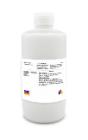
Description:
Jones Method for Kidney, Silver Method, For the staining of basement membranes, reticulum fibers, collagen, and nuclei, Stain results - Basement membranes, reticulum fibers: Black, Nuclei: Blue, Cytoplasm, collagen
Description:
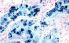
Prussian Blue Method for Hemosiderin, For the staining of deposits of hemosiderin, Stain results - Hemosiderin: Blue or Green, Nuclei: Red, Background: Pink
Description:
specialized dye for marking tissue biopsies and pathology specimens, use with frozen or fixed tissues, specially formulated binding pigment evenly coats tissue without penetrating tissue surface, fast drying, easy-to-use, and idea
Description:
Thomas Method for Malarial Parasites, Thomas (1953), Stain results - Nuclei: Blue, Plasma Cell Cytoplasm: Blue, Malarial Parasites: Blue, Erythrocytes: Pink, Other Tissue Elements: Shades of Rose to Red
Description:
Jones Method for Kidney, Silver Method, For the staining of basement membranes, reticulum fibers, collagen, and nuclei, Stain results - Basement membranes, reticulum fibers: Black, Nuclei: Blue, Cytoplasm, collagen
Description:
Thomas Method for Malarial Parasites, Thomas (1953), Stain results - Nuclei: Blue, Plasma Cell Cytoplasm: Blue, Malarial Parasites: Blue, Erythrocytes: Pink, Other Tissue Elements: Shades of Rose to Red
Description:
Puchtler-Sweat Method for Basement Membranes, Puchtler and Sweat (1964), Stain results - Basement Membranes: Black in cross section/Gray in tangential sections, Nuclei: Pink to Red
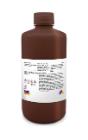
Description:
Laqueur's Method for Alcoholic Hyalin Lacqueur (1950), For mallory bodies, erythrocytes, bile pigment and proteinaceous, material in liver, Stainresults - Mallory Bodies: Bright Red, Erythrocytes: Red, Cytoplasm: Pale Brown
Description:
specialized dye for marking tissue biopsies and pathology specimens, use with frozen or fixed tissues, specially formulated binding pigment evenly coats tissue without penetrating tissue surface, fast drying, easy-to-use, and ide
Description:
Prussian Blue Method for Hemosiderin, For the staining of deposits of hemosiderin, Stain results - Hemosiderin: Blue or Green, Nuclei: Red, Background: Pink
Description:
Jones Method for Kidney, Silver Method, For the staining of basement membranes, reticulum fibers, collagen, and nuclei, Stain results - Basement membranes, reticulum fibers: Black, Nuclei: Blue, Cytoplasm, collagen
Description:
A Combined Hematoxylin and Eosin/Methenamine Silver Stain for the Histological Diagnosis of Fungi in Tissue Sections For bacterial, fungal stains. Stain results - Organism: Blue-Black Background: Rose
Description:
Bodian's Protargol Method, For neurological tissue stains: Nerve fiber and endings, Results - Nerve Fibers, Nuclei: Black, Myelinin, Muscle, Erythrocytes, Lissamine Fast Red Counterstain: Red, Background-Lissamine Fast Red
Description:
Hirano-Zimmerman Method for Nerve Cells and Fibers, Staining of nerofibrils, dendrites, axis cylinders, senile plaques, etc, Results - Neurofibrils, dendrites and axis cylinders: Black, Cytoplasm of astrocytes and cytoplasm
Description:
Thomas Method for Malarial Parasites, Thomas (1953), Stain results - Nuclei: Blue, Plasma Cell Cytoplasm: Blue, Malarial Parasites: Blue, Erythrocytes: Pink, Other Tissue Elements: Shades of Rose to Red
Description:
Puchtler-Sweat Method for Basement Membranes, Puchtler and Sweat (1964), Stain results - Basement Membranes: Black in cross section/Gray in tangential sections, Nuclei: Pink to Red
Description:
Laqueur's Method for Alcoholic Hyalin Lacqueur (1950), For mallory bodies, erythrocytes, bile pigment and proteinaceous, material in liver, Stainresults - Mallory Bodies: Bright Red, Erythrocytes: Red, Cytoplasm: Pale Brown
Description:
Jones Method for Kidney, Silver Method, For the staining of basement membranes, reticulum fibers, collagen, and nuclei, Stain results - Basement membranes, reticulum fibers: Black, Nuclei: Blue, Cytoplasm, collagen
Description:
Canaliculi and Lacunae Stain, Powers, Rasmussen and Clark (1951), For bones and teeth, Stain results - Canaliculi, lacunae, odontoblast and dentinal tubules: Bluish to purplish black
Description:
A Combined Hematoxylin and Eosin/Methenamine Silver Stain for the Histological Diagnosis of Fungi in Tissue Sections For bacterial, fungal stains. Stain results - Organism: Blue-Black Background: Rose
Description:
Puchtler-Sweat Method for Basement Membranes, Puchtler and Sweat (1964), Stain results - Basement Membranes: Black in cross section/Gray in tangential sections, Nuclei: Pink to Red
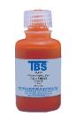
Description:
Periodic Acid Leucofuchsin Method - (PAS), For General tissue stain, Stain results - Nuclei Black on Blue, Cytoplasm Gray, Yellow or Orange, Collagen Pink, Reticulum Purplish Red, Glycogen- Dark Purplish Red
Description:
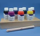
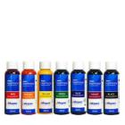
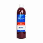
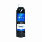
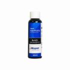
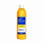
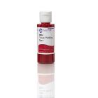
Tissue Marking Dye, Flip-Top dispenser cap, for marking margins & multiple biopsies. Dyes have been developed to define margins without bleeding, changing color or fading. Color: Red, Size: 2oz
Description:
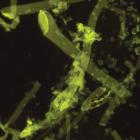
Fungi-Fluor* Kit for Fungal Detection offers a quick fluorescent stain/counterstain procedure for various fungal organisms, can be used to screen a variety of specimen types, for fungal detection, Contains: 75ml of staining solution A, 75ml of counterstaining solution B