Easy access to products and protocols for research use only in the identification of 2019-nCoV based on Centers for Disease Control and Prevention (CDC) recommendations
So much has changed during this unprecedented time, except your ability to count on Avantor. We continue to set science in motion to create a better world by providing you with the right solutions to keep moving forward.
Our solutions, developed with you as our focus, are crafted by our team and network of professionals with advanced degrees in science, quality control, engineering, manufacturing and industry experience.
Avantor supports end-to-end fluid management solutions – including peristaltic pumps and aseptic fluid transfer solutions – that are reliable and customer-centric, helping bioprocessing manufacturers meet their research and production goals.
A strong, vibrant research and development group is the lifeblood of all industries. VWR will support you from the latest life science products to the guaranteed purity of organic building blocks...
VWR is ready to support your production facility with reliable access to raw materials and essential supplies. We can also help you increase productivity...
VWR is proud of our years of experience providing choice and excellent service to the Industrial market from Food & Beverage, Petrochemical, Environmental Testing, Waste Water, Cosmetics, Consumer Goods, Agriculture and more...
VWR is your complete source for workplace supplies. Binders, calendars, pens, cleaning and sanitation supplies, and office equipment are just some of the essential products we offer...
New Avantor® J.T.Baker® premium conductive and non-conductive robotic tips deliver superior quality and reliable performance for results you can trust.
Avantor Services provides a wide range of specialized services and digital solutions to help you solve complex challenges.
We’ve built our reputation on consistent, comprehensive mastery of day-to-day operations, allowing lab, clinical, and production environments to focus their high-value resources on core scientific priorities.
As our customers’ needs have evolved, so have our capabilities. We have become experts in scientific operations, improving performance with sophisticated solutions and providing guidance on best practices.
You can select and customize services for peak efficiency, quality, and accelerated innovation.
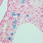
Overcome normal optical limitations in microscopic studies by highlighting specific structures with stains and reagents and then protect samples with fixatives. The fast-acting dyes create strong contrasts between cell components for easier detection or analyses. Sensitive attractions to certain organelle or cellular components differ among offered stains, allowing precise testing modifications. Flexible in use, ready-to-use stains may be handled manually or in conjunction with automatic equipment.
Description:
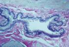
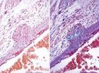
A Combined Hematoxylin and Eosin/Methenamine Silver Stain for the Histological Diagnosis of Fungi in Tissue Sections For bacterial, fungal stains. Stain results - Organism: Blue-Black Background: Rose
Description:
The PAP pen is a special marking pen that provides a thin film-like green-tinged hydrophobic barrier when a circle is drawn around a specimen on a slide.
Description:
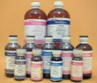
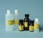
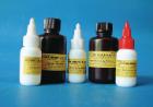
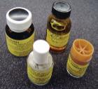
Premixed, prebuffered fixatives, pH adjusted for immediate use. Freshly prepared to ensure maximum shelf life. Fixatives are filled in 20ml glass vials (10ml/vial). This allows for the immediate fixation of your specimens
Description:
Kluver-Barrera Method for Myelin and Nerve Calls, Relation of nerve cells to neutroglia, etc, Stain results - Myelin, including phospholipids: Blue to Green cells, Cells and Cell products: Pink to Violet
Description:
CAS # N.A. Specific Gravity: 1.08-1.10 Same characteristics as Epon 812, physically, and provides same results in preservation, handling, curing and sectioning. May cause etching on selected plastics.
Description:
Stain Bluing Solution Type 1 Conc, bluing reagent concentrate, 8,0 pH to assure proper bluing of hematoxylin stained tissue sections, Resistant to carry-over and remains in the proper pH range for a long period of
Description:
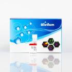
Prussian Blue Iron Stain Kit, is used to demonstrate ferric iron and ferritin, This is not a true staining technique; rather, it is a histochemical reaction, The protein is split off and allows the potassium ferrocyanide to combine with the ferric iron, size: 1 kit
Description:
Used as fixation and staining fluid. Packed in leak-proof histo-containers, prefilled, ready-to-use. Formulated with Picric Acid saturated aqueous solution 75%, Formalin (40% aqueous formaldehyde) 25% and Glacial Acetic acid 5% in de-ionized water
Description:
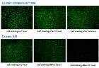
BactoView* Live Fluorescent Bacterial Stain, red fluorescent DNA-binding dye, suitable for flow cytometry or fluorescence microscopy, Stain live and dead, gram-positive and gram-negative bacteria with red fluorescence, Size: 100 ul
Description:
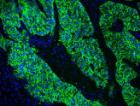
Mounting Medium with DAPI, Wet-set antifade mounting medium optimally formulated for preserving fluorescence of our CF* dyes and other fluorochromes, less viscous for rapid DAPI staining, Compatible with cyanine-based fluorophores, Size: 10 ml
Description:
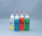
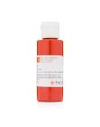
Tissue Marking Dye, Flip-Top mini bottle, Color: Black, for marking margins & multiple biopsies. Dyes have been developed to define margins without bleeding, changing color or fading. Have flip top dispenser cap, Size: 8 oz
Description:
Tissue Marking Dye* Flip-Top mini bottle, Color: Black, for marking margins and multiple biopsies. Dyes have been developed to define margins without bleeding, changing color or fading. Have flip top dispenser cap, Size: 0.5 oz
Description:
Antifadent Mountant Solutions. Medium for immunofluorescence. Designed to reduce fading of fluorescence of dyes. AF1 (Glycerol-phosphate buffered solution containing an additive for use with labeled tissue sections).
Description:
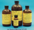
Provides excellent penetration for embedding biological tissues and rapid infiltration. Easy to prepare. Hardness is adjusted by changing proportion of flexibilizer (DER 736). Consists of: 450ml NSA, 225ml ERL 4221, 225ml DER
Description:
PCB-Free Immersion Oils for Fluorescence Microscopy. * Immersion Oil Type DF Type DF has the highest resolution of any fluorescent microscopy immersion oil
Description:
Consideration of these oils is also suggested where stage temperatures are elevated by sub-stage illuminators and high wattage projection equipment.
Description:
Lillie Modification of Masson's Trichrome, Stains cells, cytoplasm muscle and collagen of mammalian tissue, Stain results - Nuclei: Black, Cystoplasm: Brown to Pink, Muscle: Red
Description:
Tissue Marking Dye, Flip-Top mini bottle, Color: Blue, for marking margins & multiple biopsies. Dyes have been developed to define margins without bleeding, changing color or fading. Have flip top dispenser cap, Size: 8 oz
Description:
Carstairs Method for Fibrin and Platelets, For the staining of fibrin and platelets, Stain results - 48 hours or more: Fibrin: Bright Red, Platelets Gray blue to navy, Collagen: Bright Blue, Muscle: Red
Description:
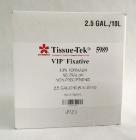
Premixed, prebuffered fixatives, pH adjusted for immediate use. Freshly prepared for maximum shelf life. Immediate fixation of specimens without transfer. 2.5% Glutaraldehyde in 0.1M Sorensen's Sodium- Potassium Phosphate B
Description:
Verhoeff's Van-Gieson's (VVG Method), For stain elastic fibers, nuclei, collagen & other tissue elements, Stain results - Elastic Fibers: Blue-black to Black-Fine elastic fibrils may not be stained with this method, Nuclei: Blue
Description:
Carstairs Method for Fibrin and Platelets, For the staining of fibrin and platelets, Stain results - 48 hours or more: Fibrin: Bright Red, Platelets Gray blue to navy, Collagen: Bright Blue, Muscle: Red, Red Blood Cells: Clear Yellow
Description:
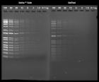
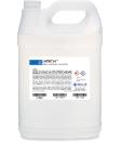
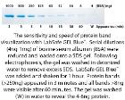
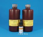
Blue 1 Gallon Cube. Enhanced protein stain that is based on Coomassie dye that offers unsurpassed sensitivity and rapid band visualization. No destaining is required; bands visualized in 10-60 minutes. Sensitivity 4-8ng will not effect transfer or other downstream applications.
Description:
Verhoeff's Van-Gieson's (VVG Method), For stain elastic fibers, nuclei, collagen & other tissue elements, Stain results - Elastic Fibers: Blue-black to Black-Fine elastic fibrils may not be stained with this method, Nuclei: Blue
Description:
Non-hazardous water-based, mounting media For mounting immunohistochemical stained tissues, which can be damaged or are soluble in organic solvents, such as xylene or toluene, Crystal/Mount allows for postmounting
Description:
Stain Alcoholic Clarifier Type 2, is designed to be used with Type 2 Hematoxylin to produce cell transparency and remove any non-specific staining, Slightly milder clarifier than the Optik Aqueous Clarifier, Size: 1
Description:
Has virtually no background fluorescence making it For observing low levels of fluorescence. Highly stable, water white, and non-hydroscopic. Can be substituted for all other fluorescence microscopy immersion oils
Description:
Thick base. Easily applied with a wooden pick. Fast drying. Average grain size less than 10um. Comes with a brush attached to the cap. Service temperature is 30 minutes at 200[degree]C.
Description:
Periodic Acid Leucofuchsin Method - (PAS), For General tissue stain, Stain results - Nuclei Black on Blue, Cytoplasm Gray, Yellow or Orange, Collagen Pink, Reticulum Purplish Red, Glycogen- Dark Purplish Red
Description:
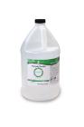
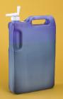
Protein Gel Stain, One-Step Lumitein, ready-to-use, aqueous-based solution that does not contain hazardous methanol or acetic acid. requires only a single 5-30 minute staining step without fixation. can detect approximately 1-10 ng of protein per band, Size: 1l
Description:
For Horizontal, Inverted and Inclined Instruments and Projection Equipment: Higher viscosities are generally required. Type NVH meets the requirement, having a viscosity of 21,000cST.
Description:
Very low viscosity (8cps), non-toxic resin. Polar monomer polyhydroxylated acromatic acrylic resin. Cured by heat or UV light. Sections of polymerized resin are hydrophilic. Consists of: 500ml LR White Resin, 10ml UV accelerator
Description:
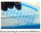
Ready to use Coomassie based protein stain. Requires no destaining or fixation step. Simple wash PAGE Gel with water, pour in stain and bands will appear without background in 10-60minutes. Final water wash will intensify bands. 4-8ng sensitivity.
Description:
Van Gieson's Method for Collagen Fibers, For collagen, muscle and cornified epithelium, Stain results - Collagen: Red, Smooth and striated muscle: Yellowish to Brownish, Cornified epithelium: Yellow, Hyalin: Yellow
Description:
Van Gieson's Method for Collagen Fibers, For collagen, muscle and cornified epithelium, Stain results - Collagen: Red, Smooth and striated muscle: Yellowish to Brownish, Cornified epithelium: Yellow, Hyalin: Yellow