Keep your session open?
Ending In 
Your shopping session has expired.
Your session has expired. For your security, we have logged you out.
Would you like to log in again?
  • Product Category
  • Criteria
  • Supplier
  • Suppliers found in search results
    Sort by:

You Searched For:

Stains

1,619  results were found
Stains

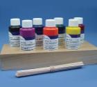
Overcome normal optical limitations in microscopic studies by highlighting specific structures with stains and reagents and then protect samples with fixatives. The fast-acting dyes create strong contrasts between cell components for easier detection or analyses. Sensitive attractions to certain organelle or cellular components differ among offered stains, allowing precise testing modifications. Flexible in use, ready-to-use stains may be handled manually or in conjunction with automatic equipment.


SearchPresentationType-SearchPresentationType-SearchPresentationType-SearchPresentationType-SearchPresentationType-SearchPresentationType-SearchPresentationType-VERTICAL


SearchResultCount:"1619"
Searching List View List View Easy View 
Sort by:


Vendor Image
Description:   
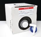
Previ* Color Gram 12, Automated Gram Staining System, Includes: User Manual, Nozzle Cleaning Soln, Empty Bottle,Waste Tubing, Waste Container, Easy To Use & Relies On An Innovative Spray Technology Which Enables Rapid, Standardized & Accurate Results For All Types Of Specimens
Catalog Number: 76204-648
Supplier: bioMérieux


Quantity:


Vendor Image
Description:   
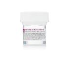
Durcupan ACM, Epoxy Resin, An aromatic polyepoxide; a colorless resin of relatively low viscosity, with virtually no shrinkage. Single Component B
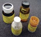
Catalog Number: 100503-854
Supplier: Electron Microscopy Sciences


Minority or Woman-Owned Business Enterprise

Quantity:


Vendor Image
Description:   Methenamine Silver Methodfor Argentaffin Cells, Staining of argentaffin granules of enterochromaffin cells, Results - Argentaffin Cells: Black, Coarse connective tissue of submucosa: Variable amount of blackening.
Catalog Number: 101411-396
Supplier: Electron Microscopy Sciences


Minority or Woman-Owned Business Enterprise

Quantity:


Product Image
Catalog Number: 76548-224
Supplier: StatLab


Quantity:


Product Image
Catalog Number: 76547-976
Supplier: StatLab


Quantity:


Vendor Image
Description:   Modified Verhoff Elastic Van Gieson Stain, For connective tissue stains - Stain with a poly-acid step, Stain results - Nuclei: Grayish-Black, Collagen Fibers: Red, Elastic Fibers: Bluish-Black, Muscle: Greenish-Yellow
Catalog Number: 100504-600
Supplier: Electron Microscopy Sciences


Minority or Woman-Owned Business Enterprise

Quantity:


Product Image
Catalog Number: 76549-734
Supplier: Mortech Manufacturing


Quantity:


Product Image
Catalog Number: 76549-750
Supplier: Mortech Manufacturing


Quantity:


Product Image
Description:   
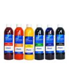
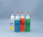
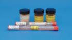
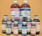
Tissue Marking Dye, Flip-Top mini bottle, Color: Gold, for marking margins & multiple biopsies. Dyes have been developed to define margins without bleeding, changing color or fading. Have flip top dispenser cap, Size: 2 oz
Catalog Number: 10809-860
Supplier: CANCER DIAGNOSTICS, INC.


Quantity:


MSDS SDS    
Product Image
Catalog Number: 76548-086
Supplier: StatLab


Quantity:


Product Image
Description:   Tissue Marking Dye, Flip-Top mini bottle, Color: Magenta, for marking margins & multiple biopsies. Dyes have been developed to define margins without bleeding, changing color or fading. Have flip top dispenser cap, Size: 2 oz
Catalog Number: 10809-914
Supplier: CANCER DIAGNOSTICS, INC.


Quantity:


MSDS SDS    
Product Image
Description:   Tissue Marking Dye, Flip-Top mini bottle, Color: Black, for marking margins & multiple biopsies. Dyes have been developed to define margins without bleeding, changing color or fading. Have flip top dispenser cap, Size: 2 oz
Catalog Number: 10809-846
Supplier: CANCER DIAGNOSTICS, INC.


Quantity:


MSDS SDS    
Vendor Image
Description:   Herxheimer's Technique, To stain fat and cholesterol, Stain results - Nuclei: Blue, Fat: Orange-Red, Cholesterol: Red
Catalog Number: 101411-404
Supplier: Electron Microscopy Sciences


Minority or Woman-Owned Business Enterprise

Quantity:


Vendor Image
Description:   Wolbach's Giemsa Method, Wolbach, Todd, and Palfrey (1922), For the staining of : nuclei, collagen, riskettsia and bacteria, Stain results - Nuclei, Bacteria: Blue, Rickettsia: Purple, Collagen
Catalog Number: 101411-542
Supplier: Electron Microscopy Sciences


Minority or Woman-Owned Business Enterprise

Quantity:


Product Image
Description:   Prussian Blue Method for Hemosiderin, Lillie (1965); Highman (1962); Perls (1867), Stain results - Hemosiderin: Blue or Green, Nuclei: Red, Background: Pink
Catalog Number: 101411-150
Supplier: Electron Microscopy Sciences


Minority or Woman-Owned Business Enterprise

Quantity:


Product Image
Description:   translucent on thin films, useful with abrasive slurry cutting of materials, suited to non-porous surfaces (glass or polished metal with a reasonably large area), soluble in a variety of solvents, Soft Hardness, Melting Point of 52°
Catalog Number: 101411-920
Supplier: Electron Microscopy Sciences


Minority or Woman-Owned Business Enterprise

Quantity:


Product Image
Description:   Lillie Modification of Masson's Trichrome, Stains cells, cytoplasm muscle and collagen of mammalian tissue, Stain results - Nuclei: Black, Cystoplasm: Brown to Pink, Muscle: Red, Erythrocytes: Brilliant Scarlet
Catalog Number: 101411-268
Supplier: Electron Microscopy Sciences


Minority or Woman-Owned Business Enterprise

Quantity:


Product Image
Description:   McManus' Method (PAS) for Glycogen, McManus (1948), Stain results - Nuclei: Blue, Fungi: Red, Background, when Light Green is used as the counter-stain: Green, pale. Refrigerated!
Catalog Number: 101411-216
Supplier: Electron Microscopy Sciences


Minority or Woman-Owned Business Enterprise

Quantity:


Product Image
Description:   
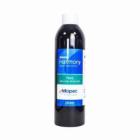
Glycerol-phosphate buffered saline based mountant solution. Addition of antifadent is to retard bleaching of fluorochromes. Particularly useful for high magnification work. Both are medium viscosity and water-clear.
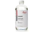
Catalog Number: 101410-704
Supplier: Electron Microscopy Sciences


Minority or Woman-Owned Business Enterprise

Quantity:


Vendor Image
Description:   Methenamine Silver Methodfor Argentaffin Cells, Staining of argentaffin granules of enterochromaffin cells, Results - Argentaffin Cells: Black, Coarse connective tissue of submucosa: Variable amount of blackening.
Catalog Number: 101411-402
Supplier: Electron Microscopy Sciences


Minority or Woman-Owned Business Enterprise

Quantity:


Product Image
Description:   Genta, Robason and Graham Stain for H-pylori & Gastric Morphology, Stain results - Muscle cells: Brownish yellow, Lamina propria ground substance: Light yellowish or grayish, Smooth muscle fibers: Light Pink
Catalog Number: 101411-062
Supplier: Electron Microscopy Sciences


Minority or Woman-Owned Business Enterprise

Quantity:


Vendor Image
Description:   Wolbach's Giemsa Method Wolbach, Todd, and Palfrey (1922) Nuclei, collagen. Stain results - Nuclei, Bacteria: Blue Rickettsia: Purple Collagen, other tissue elements: Pink to rose
Catalog Number: 101411-108
Supplier: Electron Microscopy Sciences


Minority or Woman-Owned Business Enterprise

Quantity:


Product Image
Description:   Maximow's Method for Bone Marrow, Morphology details of marrow cells, Stain results - Nuclei Blue, Basophile leukocyte, mast cell granules Purple to Violet, Cartilage Purple, Erythrocytes, eosinophile granules Pink
Catalog Number: 101411-200
Supplier: Electron Microscopy Sciences


Minority or Woman-Owned Business Enterprise

Quantity:


Product Image
Description:   
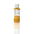
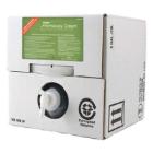
Clearium* Mounting Media, Pint, Eliminates the need for hazardous xylene or xylene substitutes as a clearing agent in histology and cytology laboratories, With Clearium, simply do away with the final step of clearing and coverslip directly from absolute isopropyl alcohol
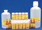
Catalog Number: 103302-098
Supplier: Electron Microscopy Sciences


Quantity:


Product Image
Description:   Conductive Gold-Paste Extender
Catalog Number: 102090-970
Supplier: Electron Microscopy Sciences


Quantity:


Product Image
Description:   Carson’s Phosphate Buffered Paraformaldehyde
Catalog Number: 102091-846
Supplier: Electron Microscopy Sciences


Quantity:


Product Image
Description:   Fluoro-Gel with Anti-Fading Agent 
Catalog Number: 102092-098
Supplier: Electron Microscopy Sciences


Quantity:


Product Image
Description:   LR White Resin, Hard, Uncatalyzed
Catalog Number: 102091-822
Supplier: Electron Microscopy Sciences


Quantity:


Product Image
Description:   Carson’s Phosphate Buffered Paraformaldehyde
Catalog Number: 102091-848
Supplier: Electron Microscopy Sciences


Quantity:


Product Image
Description:   Clear-Mount (with PIPE Buffer)
Catalog Number: 102092-132
Supplier: Electron Microscopy Sciences


Quantity:


Product Image
Description:   EMS-Mount
Catalog Number: 102092-112
Supplier: Electron Microscopy Sciences


Quantity:


Product Image
Description:   Mirsky’s Fixative Ready-to-Use
Catalog Number: 102091-834
Supplier: Electron Microscopy Sciences


Quantity:


Vendor Image
Catalog Number: 102092-802
Supplier: Electron Microscopy Sciences


Quantity:


Vendor Image
Description:   Glycerol Fluoro Mount, Color: Dark coffee, Refractive Index: 1.4563, With Para Phenylenediamine (PPD) anti-fading agent, For preserving fluorescence of tissue and cell smears, Application: Immunofluorescence, confocal microscopy, Storage: 2-8 deg C, Volume: 100ml
Catalog Number: 103306-376
Supplier: Electron Microscopy Sciences


Quantity:


Product Image
Description:   TFM* Tissue Freezing Medium, color: green, ultra pure formulation of water-soluble glycols and resins that provides a solid bond between the tissue and the object holder, freezing medium's reduced water content minimizes freeze-fracturing, capacity: 4x4 oz
Catalog Number: 100499-661
Supplier: Electron Microscopy Sciences


Quantity:


Vendor Image
Description:   Formalin Prefilled HistoTainer, 60 ml
Catalog Number: 102091-908
Supplier: Electron Microscopy Sciences


Quantity:


Product Image
Description:   
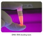
Premixed and ready to load gel dyes. Designed to minimize the use of ethidium bromide and the risk associated with the regular use of ethidium bromide. BromoBlue and no ethidium bromide, Size: 1.5mL.
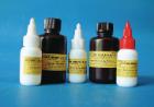
Catalog Number: 82022-462
Supplier: G-Biosciences


Quantity:


Vendor Image
Description:   is a cAMP analog and can be used to probe cAMP receptors.
Catalog Number: 89138-102
Supplier: Biotium


Quantity:


Product Image
Description:   PermaBlack/HRP is a substrate-chromogen system designed to be used for either IHC or ISH when utilizing horseradish peroxidase. PermaBlack/HRP can be permanently mounted to produce a sharply contrasting ebony black color that can be easily distinguished from other stains. 110ml.
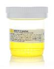
Catalog Number: 10064-834
Supplier: Diagnostic Biosystems


Quantity:


Product Image
Description:   PermaYellow/HRP is a substrate-chromogen system designed to be used for either IHC or ISH when utilizing horseradish peroxidase. PermaYellow/HRP produces a distinct bright yellow that can easily be distinguished from other stains. 110ml.
Catalog Number: 10064-852
Supplier: Diagnostic Biosystems


Quantity:


Product Image
Description:   PermaGreen Plus/AP is a substrate-chromogen system designed for either IHC or ISH when utilizing alkaline phosphatase. It shows increased staining intensity, and produces a strong green color that is insoluble in alcohol and xylene. 110ml.
Catalog Number: 10064-766
Supplier: Diagnostic Biosystems


Quantity:


Product Image
Description:   Harris Hematoxylin
Catalog Number: 102092-388
Supplier: Electron Microscopy Sciences


Quantity:


Product Image
Description:   It has a refractive index (nD at 25[degree]C) of 1.662 and an Abbe V dispersion of 26, optically similar to Aroclor 5442 but is PCB-free.
Catalog Number: 100496-580
Supplier: Electron Microscopy Sciences


Minority or Woman-Owned Business Enterprise

Quantity:


Vendor Image
Description:   offers all of the same features as JB-4 with the following additions: It produces less of an exothermic reaction than JB4 which is good for temperature sensitive tissues. It produces harder blocks which is ideal for dense samples.
Catalog Number: 100496-478
Supplier: Electron Microscopy Sciences


Minority or Woman-Owned Business Enterprise

Quantity:


Vendor Image
Description:   Electrically and thermally conductive, Silver filled. One part system, Inorganic system for bonding applications up to 1200°F
Catalog Number: 100497-408
Supplier: Electron Microscopy Sciences


Minority or Woman-Owned Business Enterprise

Quantity:


Vendor Image
Description:   A modification of Bouin's solution, stable, will decalcify small bone specimens, can be stained, cupric acetate in the solution stabilizes red blood cell membranes and cosinophil and endocrine cell granules so that less lysis occurs than with Bouin's solution
Catalog Number: 100498-614
Supplier: Electron Microscopy Sciences


Minority or Woman-Owned Business Enterprise

Quantity:


Product Image
Description:   specialized dye for marking tissue biopsies and pathology specimens, use with frozen or fixed tissues, specially formulated binding pigment evenly coats tissue without penetrating tissue surface, fast drying, easy-to-use, and ideal f
Catalog Number: 100498-296
Supplier: Electron Microscopy Sciences


Minority or Woman-Owned Business Enterprise

Quantity:


Product Image
Description:   It has a refractive index (nD at 25[degree]C) of 1.582 and an Abbe V dispersion of 33. Its optical clarity makes it the preferred choice for minimum visible absorption.
Catalog Number: 100502-412
Supplier: Electron Microscopy Sciences


Minority or Woman-Owned Business Enterprise

Quantity:


Vendor Image
Description:   Diethylene Glycol Distearate (DEGDS/DGD), a removable embedding medium (for thin and thick sections) provides embedment free sections. Good for immunolabeling and high resolution light microscopy. Kit consists of: 450ml DGD, 450ml Ethanol, 450ml n-Butyl alcohol
Catalog Number: 100503-430
Supplier: Electron Microscopy Sciences


Minority or Woman-Owned Business Enterprise

Quantity:


Product Image
Description:   Modified Steiner - Using Chapman's Modification For bacterial stain. Stain results - Spirochetes, Donovan Bodies, General Bacteria,Legionairies Disease Bacteria: Dark Brown or Black Background: Bright Yellow to light brown
Catalog Number: 100504-536
Supplier: Electron Microscopy Sciences


Minority or Woman-Owned Business Enterprise

Quantity:


Vendor Image
Description:   A modification of Bouin's solution, stable, will decalcify small bone specimens, can be stained, cupric acetate in the solution stabilizes red blood cell membranes and cosinophil and endocrine cell granules so that less lysis occurs than with Bouin's solution
Catalog Number: 100502-582
Supplier: Electron Microscopy Sciences


Minority or Woman-Owned Business Enterprise

Quantity:


Vendor Image
Description:   May-Grunwald / Giemsa Combined Stain, Beck (1938); Pappanheim (1912), Stain results - Nuclei, Bacteria Blue, Cytoplasm Pink-Rose
Catalog Number: 101411-192
Supplier: Electron Microscopy Sciences


Minority or Woman-Owned Business Enterprise

Quantity:


Vendor Image
Description:   Gomori's Methenamine Silver for Urate Crystals Urate crystals, Stain results - Urate Crystals: Black, Background: Green
Catalog Number: 101411-448
Supplier: Electron Microscopy Sciences


Minority or Woman-Owned Business Enterprise

Quantity:


MSDS SDS    
Vendor Image
Description:   Methenamine Silver Methodfor Argentaffin Cells, Staining of argentaffin granules of enterochromaffin cells, Results - Argentaffin Cells: Black, Coarse connective tissue of submucosa: Variable amount of blackening.
Catalog Number: 101411-390
Supplier: Electron Microscopy Sciences


Minority or Woman-Owned Business Enterprise

Quantity:


Vendor Image
Description:   Wilder Modification of Bielschowsky's Method, Staining of Collagen and reticulum, results - Reticulum Fibers (If beading observed and/or dark background, replace ammonium hydroxide silver, Nitrate solution & reducing solution)
Catalog Number: 101411-340
Supplier: Electron Microscopy Sciences


Minority or Woman-Owned Business Enterprise

Quantity:


Product Image
Description:   Jones Method for Kidney, Silver Method, For the staining of basement membranes, reticulum fibers, collagen, and nuclei, Stain results - Basement membranes, reticulum fibers: Black, Nuclei: Blue, Cytoplasm, collagen
Catalog Number: 101411-352
Supplier: Electron Microscopy Sciences


Minority or Woman-Owned Business Enterprise

Quantity:


Vendor Image
Description:   Gomori's Methenamine Silver for Urate Crystals Urate crystals, Stain results - Urate Crystals: Black, Background: Green
Catalog Number: 101411-458
Supplier: Electron Microscopy Sciences


Minority or Woman-Owned Business Enterprise

Quantity:


Vendor Image
Description:   Woelcker's Method for Myelin Sheath, Stain for myelin sheath, glial cells and nucleoli of neurons, Stain results - Glial Cells, Nucleoli of neurons: Black, Myelin sheath: Blue, Background: Clear
Catalog Number: 101411-484
Supplier: Electron Microscopy Sciences


Minority or Woman-Owned Business Enterprise

Quantity:


Vendor Image
Description:   The LumiGLO Western Blot Kit provides the combination of a highly specific, stable liquid conjugate with a sensitive chromogenic substrate allows rapid and accurate identification of samples. All solutions required for detection are provided.
Catalog Number: 95059-302
Supplier: KPL


Quantity:


MSDS SDS    
Product Image
Catalog Number: 76579-378
Supplier: Mortech Manufacturing


Quantity:


Product Image
Description:   AdvanStain Scarlet Kit
Catalog Number: 490015-238
Supplier: Advansta


Quantity:


Product Image
Description:   
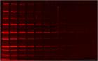
is a luminescent dye designed for detecting total proteins in polyacrylamide gels following gel electrophoresis. The stain combines excellent sensitivity, exceptional user-friendliness and compatibility. Ready to use formulation.
Catalog Number: 89138-836
Supplier: Biotium


Quantity:


Product Image
Description:   Immersion Oil Type OVH
Catalog Number: 102092-090
Supplier: Electron Microscopy Sciences


Quantity:


Product Image
Description:   PAGE GelRed* is a non-toxic, non-mutagenic red DNA gel stain specifically designed to stain DNA in polyacrylamide gels. PAGE GelRed* can be imaged using a 254 nm UV transilluminator with an ethidium bromide filter. Supplied as a ready-to-use 1X gel staining solution in water.
Catalog Number: 10154-218
Supplier: Biotium


Safer Human and Environmental Health

Quantity:


MSDS SDS    
Items Per Page: 16  32  64 
833 - 896  of 1,619
Prev   14  15  16  17  18  19  20  21  22  23  24  25  26  Next