Easy access to products and protocols for research use only in the identification of 2019-nCoV based on Centers for Disease Control and Prevention (CDC) recommendations
So much has changed during this unprecedented time, except your ability to count on Avantor. We continue to set science in motion to create a better world by providing you with the right solutions to keep moving forward.
Our solutions, developed with you as our focus, are crafted by our team and network of professionals with advanced degrees in science, quality control, engineering, manufacturing and industry experience.
Avantor supports end-to-end fluid management solutions – including peristaltic pumps and aseptic fluid transfer solutions – that are reliable and customer-centric, helping bioprocessing manufacturers meet their research and production goals.
A strong, vibrant research and development group is the lifeblood of all industries. VWR will support you from the latest life science products to the guaranteed purity of organic building blocks...
VWR is ready to support your production facility with reliable access to raw materials and essential supplies. We can also help you increase productivity...
VWR is proud of our years of experience providing choice and excellent service to the Industrial market from Food & Beverage, Petrochemical, Environmental Testing, Waste Water, Cosmetics, Consumer Goods, Agriculture and more...
VWR is your complete source for workplace supplies. Binders, calendars, pens, cleaning and sanitation supplies, and office equipment are just some of the essential products we offer...
New Avantor® J.T.Baker® premium conductive and non-conductive robotic tips deliver superior quality and reliable performance for results you can trust.
Avantor Services provides a wide range of specialized services and digital solutions to help you solve complex challenges.
We’ve built our reputation on consistent, comprehensive mastery of day-to-day operations, allowing lab, clinical, and production environments to focus their high-value resources on core scientific priorities.
As our customers’ needs have evolved, so have our capabilities. We have become experts in scientific operations, improving performance with sophisticated solutions and providing guidance on best practices.
You can select and customize services for peak efficiency, quality, and accelerated innovation.
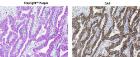
Overcome normal optical limitations in microscopic studies by highlighting specific structures with stains and reagents and then protect samples with fixatives. The fast-acting dyes create strong contrasts between cell components for easier detection or analyses. Sensitive attractions to certain organelle or cellular components differ among offered stains, allowing precise testing modifications. Flexible in use, ready-to-use stains may be handled manually or in conjunction with automatic equipment.
Description:
Resin, Lowicryl resins are highly cross-linked acrylate-based embedding resins, designed for use over a wide range of embedding conditions. These resins provide low viscosity at low temperatures. The K4M kit is usable to -35[degree]C
Description:
Resin, Lowicryl resins are highly cross-linked acrylate-based embedding resins, designed for use over a wide range of embedding conditions. These resins provide low viscosity at low temperatures.
Description:
Taylor's Method for Gram Positive and Gram Negative Bacteria, Stain results - Gram-Positive Organism: Blue to Blue-Black, Gram Negative Organism: Bright Red, Nuclei: Brownish Red, Erythrocytes: Red to Yellow-Green
Description:
Durcupan, Water Soluble, A water soluble embedding medium for EM based on an aliphatic polyepoxide. A Fluka A.G. Buchs. Switzerland-Registered Trademark. Single Component A
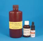
Description:
Histocryl acrylic resin specially formulated for light microscopy. Water clear. It is hydrophilic (polar) permits use of routine staining techniques without prior removal or etching. Consists of: 500ml Histocryl Resin, 50g Histocryl Catalyst (B.P.), 10ml Histocryl Accelerator
Description:
Weigert's Iron Hematoxylinwith Methachromic Dyes, For Nuclear stain, Stain results - Nuclei Black, Cytoplasm Gray-Green, Mucus, Cartilage and Yellowish Brown, Deep Red or, Mast Cell Granules Orange-Red depending on dye used
Description:
Periodic Acid Leucofuchsin Method - (PAS), For General tissue stain, Stain results - Nuclei Black on Blue, Cytoplasm Gray, Yellow or Orange, Collagen Pink, Reticulum Purplish Red, Glycogen- Dark Purplish Red
Description:
Mayer Mucicarmine Method, For mucosubstances stains, Note: rose color due to carmin staining will be obscured if sections are overstained with Weigert's Hematoxylin and/or Metanil Yellow solution.
Description:
Alizarin Red S and Toluidine Blue O, Williams (1941); Dawson (1926), Distinction between bone and cartilage in mammalian embryos, Stain results - Soft Tissues: Transparent, Osseous Tissues: Deep Red, Cartilage: Dark Blue
Description:
Mallory-Heidenhain Azan-Gomori's Modification for Islet Cells, For staining of alpha, beta and D-cells of islets of langerhans, Stain results - (Bouin Fixation), Human Tissue, Alpha granules: Red, bright
Description:
offers all of the same features as JB-4 with the following additions: It produces less of an exothermic reaction than JB4 which is good for temperature sensitive tissues. It produces harder blocks which is ideal for dense samples.
Description:
Bodian's Protargol Method, For neurological tissue stains: Nerve fiber and endings, Results - Nerve Fibers, Nuclei: Black, Myelinin, Muscle, Erythrocytes, Lissamine Fast Red Counterstain: Red, Background-Lissamine Fast Red
Description:
Modified Alizarin Red S for Fetal Specimens, Cumley, Crow, and Griffin (1939), For Minute bones and fetal ossification in mammalian embryos, Stain results - Bone: Red, Soft Tissue: . Transparent and unstained
Description:
Dane's Method for Prekeratin, Keratin, and Mucin, Keratin stain method for the staining of keratin, prekeratin, acid mucopolysaccharides and nuclei, Stain results - Acid Mucopolysaccharides: Blue, Prekeratin and Keranin: Orange to Red
Description:
Mallory-Heidenhain Azan-Gomori's Modification for Islet Cells, For staining of alpha, beta and D-cells of islets of langerhans, Stain results - (Bouin Fixation), Human Tissue, Alpha granules: Red, bright
Description:
Van Gieson's Method for Collagen Fibers, For collagen, muscle and cornified epithelium, Stain results - Collagen: Red, Smooth and striated muscle: Yellowish to Brownish, Cornified epithelium: Yellow, Hyalin: Yellow
Description:
Laqueur's Method for Alcoholic Hyalin Lacqueur (1950), For mallory bodies, erythrocytes, bile pigment and proteinaceous, material in liver, Stainresults - Mallory Bodies: Bright Red, Erythrocytes: Red, Cytoplasm: Pale Brown, Bile
Description:
Modified Giemsa Stain Kit For Quick Turn Around Results For Rapid Blood Smears For Differential Assessment And Detection Of H. Pylori Microorganisms. Complete With A Fixative For Air Dried Cell Suspensions
Description:
Modified Giemsa Stain Kit For Quick Turn Around Results For Rapid Blood Smears For Differential Assessment And Detection Of H. Pylori Microorganisms. Complete With A Fixative For Air Dried Cell Suspensions
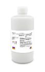
Description:
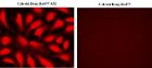
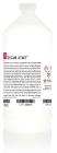
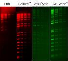
Fluorescent total protein stain kit, TotalStain Q (NC) is a fluorescent total protein stain for nitrocellulose membranes, sufficient for 15 mini-blots, Compatible with chemiluminescent and NIR fluorescence Western Blot, Size: 465ml
Description:
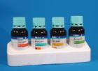
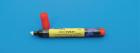
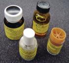
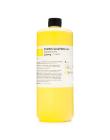
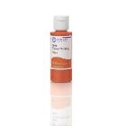
Tissue Marking Dye, Flip-Top mini bottle, Color: Orange, for marking margins & multiple biopsies. Dyes have been developed to define margins without bleeding, changing color or fading. Have flip top dispenser cap, Size: 2 oz
Description:
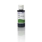
Tissue Marking Dye, Flip-Top mini bottle, Color: Green, for marking margins & multiple biopsies. Dyes have been developed to define margins without bleeding, changing color or fading. Have flip top dispenser cap, Size: 8 oz
Description:
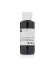
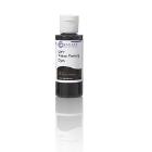
Tissue Marking Dye, Flip-Top dispenser cap, for marking margins & multiple biopsies. Dyes have been developed to define margins without bleeding, changing color or fading. Color: Black, Size: 2oz
Description:
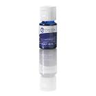
Tissue Marking Dye, Flip-Top mini bottle, Color: Blue, for marking margins & multiple biopsies. Dyes have been developed to define margins without bleeding, changing color or fading. Have flip top dispenser cap, Size: 0.5 oz
Description:
PMA Enhancer for Gram Negative Bacteria was designed to improve PMA-mediated discrimination between live and dead gram-negative bacteria. PMA Enhancer is provided as a 5X solution, and is added to a sample before the addition of PMA.
Description:
It is a two component, 100% solid silver-filled epoxy system, silver-resin paste and liquid hardener, mixing ration is 1:1. Features high thermal conductivity, and is very well suited for extensive high temperature applications (300 – 400degC)