Easy access to products and protocols for research use only in the identification of 2019-nCoV based on Centers for Disease Control and Prevention (CDC) recommendations
So much has changed during this unprecedented time, except your ability to count on Avantor. We continue to set science in motion to create a better world by providing you with the right solutions to keep moving forward.
Our solutions, developed with you as our focus, are crafted by our team and network of professionals with advanced degrees in science, quality control, engineering, manufacturing and industry experience.
Avantor supports end-to-end fluid management solutions – including peristaltic pumps and aseptic fluid transfer solutions – that are reliable and customer-centric, helping bioprocessing manufacturers meet their research and production goals.
A strong, vibrant research and development group is the lifeblood of all industries. VWR will support you from the latest life science products to the guaranteed purity of organic building blocks...
VWR is ready to support your production facility with reliable access to raw materials and essential supplies. We can also help you increase productivity...
VWR is proud of our years of experience providing choice and excellent service to the Industrial market from Food & Beverage, Petrochemical, Environmental Testing, Waste Water, Cosmetics, Consumer Goods, Agriculture and more...
VWR is your complete source for workplace supplies. Binders, calendars, pens, cleaning and sanitation supplies, and office equipment are just some of the essential products we offer...
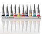
New Avantor® J.T.Baker® premium conductive and non-conductive robotic tips deliver superior quality and reliable performance for results you can trust.
Avantor Services provides a wide range of specialized services and digital solutions to help you solve complex challenges.
We’ve built our reputation on consistent, comprehensive mastery of day-to-day operations, allowing lab, clinical, and production environments to focus their high-value resources on core scientific priorities.
As our customers’ needs have evolved, so have our capabilities. We have become experts in scientific operations, improving performance with sophisticated solutions and providing guidance on best practices.
You can select and customize services for peak efficiency, quality, and accelerated innovation.
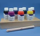
Overcome normal optical limitations in microscopic studies by highlighting specific structures with stains and reagents and then protect samples with fixatives. The fast-acting dyes create strong contrasts between cell components for easier detection or analyses. Sensitive attractions to certain organelle or cellular components differ among offered stains, allowing precise testing modifications. Flexible in use, ready-to-use stains may be handled manually or in conjunction with automatic equipment.
Description:
specifically formulated for use in microscope slide mounting and optical coupling applications, Has a refractive index (nD @25[degree]C) of 1.539 and an Abbe V dispersion of 45, optically similar to Canada Balsam
Description:
A modification of Bouin's solution, stable, will decalcify small bone specimens, can be stained, cupric acetate in the solution stabilizes red blood cell membranes and cosinophil and endocrine cell granules so that less lysis occurs than with Bouin's solution
Description:
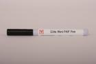
specialized dye for marking tissue biopsies and pathology specimens, use with frozen or fixed tissues, specially formulated binding pigment evenly coats tissue without penetrating tissue surface, fast drying, easy-to-use, and ide
Description:
Periodic Acid Leucofuchsin Method - (PAS), For General tissue stain, Stain results - Nuclei Black on Blue, Cytoplasm Gray, Yellow or Orange, Collagen Pink, Reticulum Purplish Red, Glycogen- Dark Purplish Red
Description:
Adhesive and sealant for use with glass, non plastic and other non-reactive materials. Spreads quickly and evenly. Fast-drying (20 minutes). Refractive index close to glass (1.510). Chemically neutral. No reaction to staining.
Description:
Rapid mounting media for microscopy. Long life preparations, without bubble formation at high ambient temperatures. For all dehydrated microscopic preparations. Cure time - 20 min at room temp. Colorless with an acid number less than 2.50
Description:
A Combined Hematoxylin and Eosin/Methenamine Silver Stain for the Histological Diagnosis of Fungi in Tissue Sections For bacterial, fungal stains. Stain results - Organism: Blue-Black Background: Rose
Description:
Bodian's Protargol Method, For neurological tissue stains: Nerve fiber and endings, Results - Nerve Fibers, Nuclei: Black, Myelinin, Muscle, Erythrocytes, Lissamine Fast Red Counterstain: Red, Background-Lissamine Fast Red
Description:
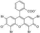
Giemsa Thick Film Stain, Lillie (1965); Barber and Komp (1929), Stain malarial parasites, Stain results - Malarial Parasites: Clear Red Chromatin, Cytoplasm: Clear Blue, Red Corpuscles: Not seen due to lacking of
Description:
Jones Method for Kidney, Silver Method, For the staining of basement membranes, reticulum fibers, collagen, and nuclei, Stain results - Basement membranes, reticulum fibers: Black, Nuclei: Blue, Cytoplasm, collagen
Description:
Canaliculi and Lacunae Stain, Powers, Rasmussen and Clark (1951), For bones and teeth, Stain results - Canaliculi, lacunae, odontoblast and dentinal tubules: Bluish to purplish black
Description:
Hirano-Zimmerman Method for Nerve Cells and Fibers, Staining of nerofibrils, dendrites, axis cylinders, senile plaques, etc, Results - Neurofibrils, dendrites and axis cylinders: Black, Cytoplasm of astrocytes and cytoplasm
Description:
Jones Method for Kidney, Silver Method, For the staining of basement membranes, reticulum fibers, collagen, and nuclei, Stain results - Basement membranes, reticulum fibers: Black, Nuclei: Blue, Cytoplasm, collagen
Description:
Puchtler-Sweat Method for Basement Membranes, Puchtler and Sweat (1964), Stain results - Basement Membranes: Black in cross section/Gray in tangential sections, Nuclei: Pink to Red
Description:
Laqueur's Method for Alcoholic Hyalin Lacqueur (1950), For mallory bodies, erythrocytes, bile pigment and proteinaceous, material in liver, Stainresults - Mallory Bodies: Bright Red, Erythrocytes: Red, Cytoplasm: Pale Brown
Description:
For Normal Light Microscopy - The greater the gap between cover glass and objective, or condenser and slide, the more desirable high viscosity becomes. For extremely large gaps, type NVH should be used.
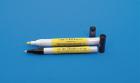
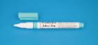
Description:
Instant conductive silver traces. Valve tip allows very smooth flow with normal writing pressure. Spring loaded. For conductivity traces, solderable termination's are possible. Tin, lead, or silver solder can be used. Silver content
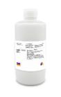
Description:
Stain Aqueous Clarifier Type 1, Clarifier is a uniquely formulated buffer that enhances cytoplasmic details, increases cell transparency and helps eliminate background staining, Convenient and ready-to-use, Size: 1 gal
Description:
Kluver-Barrera Method for Myelin and Nerve Calls, Relation of nerve cells to neutroglia, etc, Stain results - Myelin, including phospholipids: Blue to Green cells, Cells and Cell products: Pink to Violet
Description:
For microscopy. A mixture of Distyrene, a plasticizer, and xylene. A colorless synthetic resin mounting media which replaces Xylene-Balsam. It preserves the stain and dries quickly.
Description:
Stain Bluing Solution Type 1, ready-to-use, buffered bluing reagent, 8.0 pH to assure proper bluing of hematoxylin stained tissue sections, Resistant to carry-over and remains in the proper pH range for an extended time,
Description:
Acrytol, rapid-drying mounting medium, low viscosity, prevents air bubbles, contains an antioxidant to inhibit stain fading and prevent the formulation of annual rings, 1Pint
Description:
Very low viscosity (8cps), non-toxic resin. Polar monomer polyhydroxylated acromatic acrylic resin. Cured by heat or UV light. Sections of polymerized resin are hydrophilic. Consists of: 500ml LR White Resin, 10ml UV accelerator
Description:
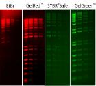
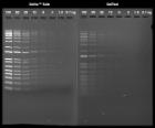
A rapid and reversible protein stain for transfer membranes. Procedure takes 10 minutes and works with nitrocellulose and PVDF staining protein bands and leaving background white. Sensitivity 2ng. Destain membrane by rinsing in warm water 10 minutes. 25 Blots.
Description:
excellent adhesion to metals, glass, and ceramic, for precise high purity work, leaves no residue after dissolving, does not clog the diamond wheel
Description:
Glycerol Mounting Medium, for immunofluorescence, This is an aqueous mounting medium made with glycerol is for preserving fluorescence of tissue and cell smears, Prevents rapid photobleaching of FITC, Texas Red, AMCA, Cy dyes, Alexa fluoro 488, size: 30 ml
Description:
Very low viscosity (8cps), non-toxic resin. Polar monomer polyhydroxylated acromatic acrylic resin. Cured by heat or UV light. Sections of polymerized resin are hydrophilic. Consists of: 500ml LR White Resin, 10ml UV accelerator, LR
Description:
Tissue Marking Dye, Applicator Series*, Brush Tip Applicator designed for small biopsies where precise application is required, Color: Blue, Size: 10oz
Description:
Stain Aqueous Clarifier Type 1 Con, Uniquely formulated buffer to enhance cytoplasmic details, increas cell transparency and help eliminate background staining, Concentrated solution to be mixed, Size: 500ml bottl
Description:
A mixture of xylene - mixed isomers and non hazardous ingredients to offer a very good synthetic mountant. Once dried the refractive index is 1.58.